【人気ダウンロード!】 heterogeneous echotexture uterus radiology 346257-What does heterogeneous echotexture of the uterus mean
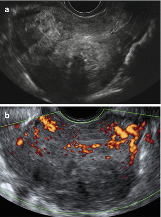
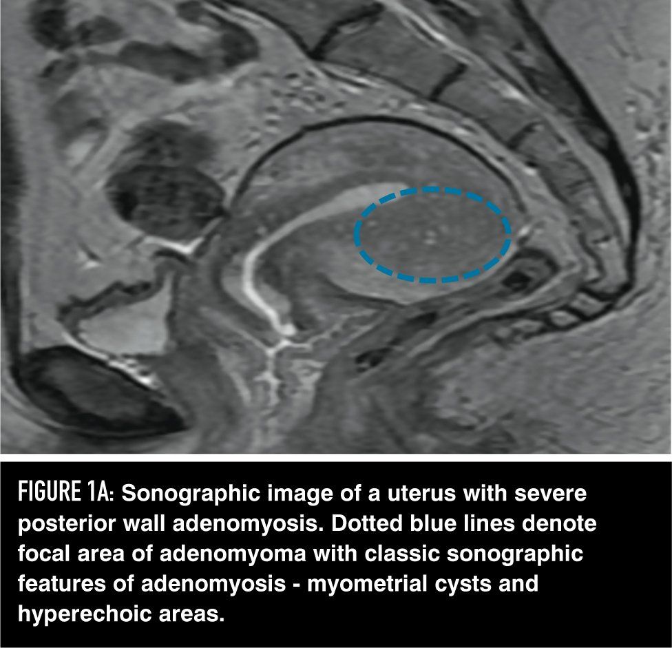
Endometrial carcinoma (EC) is the most common gynecologic malignancy in the United States Prognosis depends on patient age, histological grade, depth of myometrial invasion and/or cervical invasion, and the presence of lymph node metastases Although EC is Adenomyosis was diagnosed when a poorly defined area of abnormal echotexture (decreased or increased echogenicity, heterogeneous echotexture, myometrial cysts) was present in the myometrium All endovaginal US findings were correlated with those from histologic examination RESULTS Endovaginal US depicted 25 of 29 pathologically proved cases ofAdenomyosis of the uterus this disease is characterized by the proliferation of cells in the muscle layer of the uterus The ultrasound study clearly shows that the structure is heterogeneous This indicates that black, round neoplasms a cyst have appeared in the uterine muscle layer This leads to periodic changes in the size of the uterus
When Would You Suspect Fibroid Uterus
What does heterogeneous echotexture of the uterus mean
What does heterogeneous echotexture of the uterus mean- A heterogeneous uterus is a term used to describe the appearance of the uterus after an ultrasound is conducted It simply means that the uterus is not totally uniform in appearance during the ultrasound According to MedHelp, there are two common causes of a heterogeneous uterus uterine fibroids and adenomyosis The echotexture of the nodule was categorized as homogeneous or heterogeneous echotexture based on the uniformity of nodule echogenicity (Fig 3) Heterogeneous echotexture was determined when the solid component of a nodule showed two obviously different portions of echogenicity (isoechoic or hyperechoic vs any degree of hypoechogenicity) The echogenicity of nodules with heterogeneous


Adenomyosis A Sonographic Diagnosis Radiographics
A heterogeneous myometrial echotexture on ultrasound is usually a nonspecific finding, although it has been described with uterine adenomyosisHeterogeneous refers to a structure with dissimilar components or elements, appearing irregular or variegated For example, a dermoid cyst has heterogeneous attenuation on CT It is the antonym for homogeneous, meaning a structure with similar components Heterogenous refers to a structure having a foreign origin For example, heterogenous bone formation is bone where bone should not exist To make matters worse, heterogenous bone formation is often also heterogeneous! Coarsened hepatic echotexture is a sonographic descriptor where there uniform smooth hepatic echotexture of the liver is lost This can occur due to number of reasons which include conditions that cause hepatic fibrosis 1 cirrhosis hemochromatosis Click to see full answer Accordingly, what causes a coarse liver?
Echotexture varies from hypoechoic to hyperechoic relative to the surrounding subcutaneous fat Pure fat is essentially free of echoes Although leiomyomas are uncommon outside the uterus and gastrointestinal tract, they may be found in the extremities and take one of three forms cutaneous leiomyoma (most common, located in the dermis), angioleiomyoma"what does heterogeneous echotexture of the liver mean?When an ultrasound examination of the liver is done sound waves are sent through the liver and the transducer picks up the reflected waves to generate a picture In a healthy liver the tissue is fairly constant all throughout the organ It generat
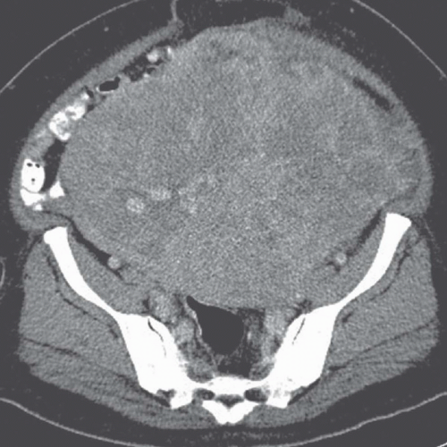
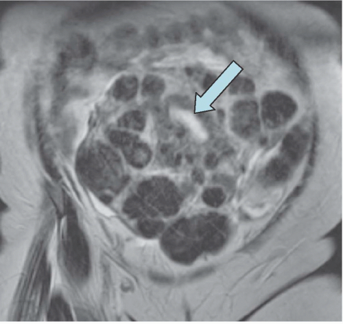
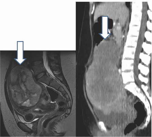
My liver ultrasound shows normal size and contour with coarsened, hyperechoic and heterogeneous echotexture without obscuration of the portal triads ?Dr Michael Gabor answered 33 years experience Diagnostic Radiology It means the texture of the uterus is not uniform on ultrasound A heterogeneous myometrial echotexture on ultrasound can be a non specific finding, of no sign Read MoreSagittal T2W MRI image shows an enlarged heterogeneous uterus containing multiple nodules (arrows) Hysterectomy and histology showed that this was diffuse leiomyomatosis There was no evidence of extrauterine spread Lipoleiomyomas Lipoleiomyomas Figure 15 are rare fatcontaining fibroids, with a reported prevalence of between 0005 and 02% They are benign and


Normal Uterus On Endovaginal Sonography The Normal Myometrium M Is Download Scientific Diagram


What Are The Most Reliable Signs For The Radiologic Diagnosis Of Uterine Adenomyosis An Ultrasound And Mri Prospective Sciencedirect
Dr Michael Gabor answered Diagnostic Radiology 33 years experience (a) Sagittal transabdominal US image shows the uterus (*), which is enlarged and heterogeneous due to fibroids (b) On a transverse transabdominal US image obtained with a 6MHz curvedarray transducer and harmonic imaging, the right ovary is displaced superior and lateral to the uterus in a superficial location The left ovary (not shown) was Equally important to the thickening is the altered echotexture of the myometrium, which becomes heterogeneous and coarse, with thin vertical shadows, resulting in a venetian blind appearance (Fig 4b)
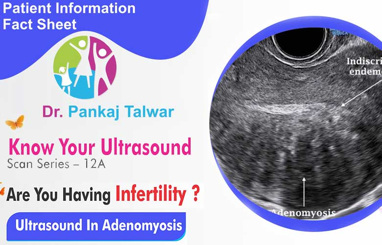


Ultrasound In Adenomyosis Fertility Treatment Center Delhi Ncr
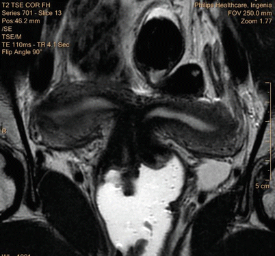


Benign Disease Of The Uterus Springerlink
Menu Close Back HomeWhat does a heterogeneous echotexture uterus mean Feb 07, The gold standard to evaluate the uterine cavity is a hysteroscopy Heterogeneous myometrial echotexture Heterogeneous myometrial echotexture Dr Daniel J Bell and Dr Yuranga Weerakkody et al, From Gorrie et al, 25, Layers of myometrium showing the three layers of smooth muscle fiber, See more resultsHeterogeneous Echotexture Definition 1 A description of heterogeneous density elements seen in a tissue composition image obtained by sonography (NCI Thesaurus) Definition 2 The breast texture is characterized by multiple small areas of increased and decreased echogenicity (NCI Thesaurus/DICOM)
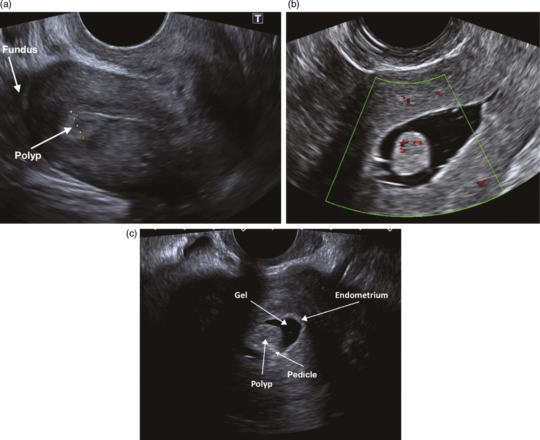


Ultrasound Imaging Of Women With Abnormal Uterine Bleeding Aub Chapter 13 Ultrasound In Reproductive Healthcare Practice


1
What Is a Heterogeneous Liver?Heterogeneous echogenicity of the thyroid gland significantly lowers the specificity, PPV, and accuracy of US in the differentiation of thyroid nodules Therefore, caution is required during evaluation of thyroid nodules detected in thyroid parenchyma showing heterogeneousHeterogeneous echogenicity of the thyroid gland is a nonspecific finding and is associated with conditions diffusely affecting the thyroid gland These


Preimplantation 3d Ultrasound Current Uses And Challenges


Principles And Clinical Uses Of Real Time Ultrasonography In Female Swine Reproduction Document Gale Academic Onefile
My ulta sound also said for liver no mass and normal size" Answered by Dr Michael Gabor It means that the liver texture is not uniform Most commonly this i Two reasons "The two most common causes of heterogenous uterus are uterine fibroids, which are benign muscular growths in the uterine wall, and adenomyosis, which is a proliferation of the normal uterine glands into the muscular wall of the uterus These conditions are very common, affecting up to maybe 50% of women by middle age"Heterogeneous myometrium is more prevalent and consistent indicator for the diagnosis of adenomyosis by ultrasound, fibroids and sometimes hyperplasia, Small amount of fluid might be in the tube or simply a cyst, Pretty much cysts on your uterine wall what does a heterogeneous echotexture uterus mean Dr, Additional diagnostics required


Adenomyosis Common And Uncommon Manifestations On Sonography And Magnetic Resonance Imaging Chopra 06 Journal Of Ultrasound In Medicine Wiley Online Library


Adenomyosis A Sonographic Diagnosis Radiographics
'Heterogenous uterus' is a description used to describe the appearance of the uterus after an ultrasound exam is done All that this means is that the ultrasound appearance ofA heterogeneous liver appears to have different masses or structures inside it when imaged via ultrasound These masses may be benign genetic differences or a result of liver disease In most cases, a finding of heterogeneous liver is followed by further medical testing to determine the cause of the heterogeneity Heterogeneous myometrium is more prevalent and consistent indicator for the diagnosis of adenomyosis by ultrasound Though enlarged uterus is almost invariably present in cases of adenomyosis (93%), yet it is totally nonspecific and is also present in multiparous females (see Figure 3, Figure 4) Download Download fullsize image;


Academic Oup Com Humupd Article Pdf 4 4 337 Pdf


Q Tbn And9gctslmjxpgtqtgbl1uxknivh3sumrgnywxebtsjy6v3t Yjuo6ps Usqp Cau
29 Zeilen Enlarged, globular uterus with diffusely heterogeneous echotexture of the myometrium and small myometrial cysts Characteristic saccular contrast material collections (arrowheads) protruding beyond the normal contour of the endometrial cavity Enlarged uterus with posterior thickening of the myometrium and multiple small cystic areas (arrows)LIVER ECHOTEXTURE CONCLUSION When characterizing liver echotexture on US, the use of the spleen as an internal comparison improves interpretation consensus and confidence in Novice and Intermediate level radiology residents as demonstrated in this preliminary study Also, a tutorial to demonstrate how to apply this principle is usefulBackground According to the American College of Radiology (ACR) Breast Imaging Reporting and Data System (BIRADS), background echotexture in breast ultrasound (US) can be categorized as homogeneous or heterogeneous Purpose To prospectively evaluate the interobserver agreement of a fourcategory classification in background echotexture assessments of breast US and to


When Would You Suspect Fibroid Uterus


Complete Hydatidiform Mole Transversal Sonogram Image A Of The Download Scientific Diagram
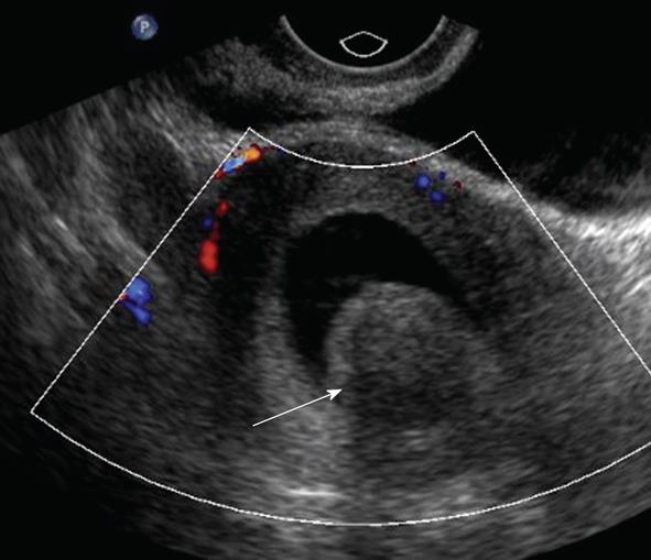
Fibroids visualized with sonography may cause obvious distortion of the uterine contour, generalized enlargement of the uterus, an altered echotexture, hypoechoic or hyperechoic focal masses, calcifications with shadowing, or heterogeneous masses with cystic areas (usually within degenerating fibroids) Fibroids are usually multiple Documentation should The heterogeneous appearance of the myometrium includes uterine enlargement and asymmetry of the anterior or posterior myometrial wall The presence of myometrial cysts (in up to 50% of cases) is highly specific for adenomyosis (,29) (,Fig 12)Heterogeneous testicular echotexture Dr Bruno Di Muzio and Dr Yuranga Weerakkody et al,PDFhe appearance of heterogeneous testes on sonography is not a rare finding in middleaged or elderly men referred for evaluation of scrotal pain, 67 years experience Endocrinology, A 54yearold female asked The uterus shows heterogenous echotexture and is diffusely
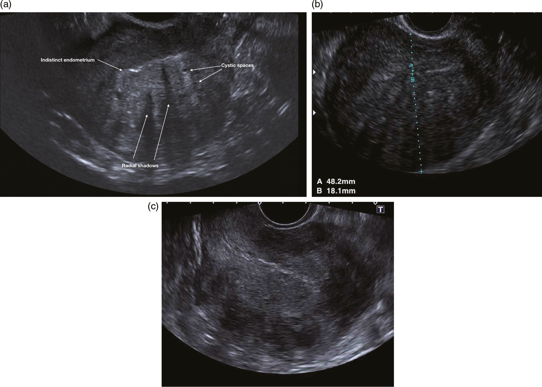


Ultrasound Imaging Of Women With Abnormal Uterine Bleeding Aub Chapter 13 Ultrasound In Reproductive Healthcare Practice


Management Of Adenomyosis Endovascular Today
Uterusconserving therapy is possible in cases of leiomyoma, whereas hysterectomy is the definitive treatment for debilitating adenomyosis Second, imaging is performed to determine the extent and depth of myometrial penetration Symptoms have been shown to correlate with the extent of disease Determining the depth of myometrial penetration is important for treatment Myometrium is the muscular wall of the uterus or it is also called as the middle layer of the uterine wall The normal condition of a uterus is called as homogeneous myometium In other words we can say that, a homogeneous myometrium is free of Lumps, voids which is the normal condition of the uterus5 Department of Radiology, EDa Hospital and IShou University, Results The accuracies of the proposed system for the classification of heterogeneous and homogeneous echotexture patterns were 9348% (43/46) and 9259% (150/162), respectively, with an overall Az (area under the receiver operating characteristic curve) of The agreement between the radiologists and


Adenomyosis Common And Uncommon Manifestations On Sonography And Magnetic Resonance Imaging Chopra 06 Journal Of Ultrasound In Medicine Wiley Online Library


Radiology Gallery Facebook
It may be heterogeneous or more echogenic than myometrium;Hypoechoic myometrial striations have been described as well There may be very small cysts in the abnormal regions, along with small echogenicHeterogeneous or mottled testes in middleaged or elderly men are often encountered on sonography To determine the prevalence, cause, and significance of this find ing, we examined 50 testes (25 pairs) from autopsy specimens with sonography and gross and microscopic pathology SUBJECTS AND METHODS Testicles were obtained at autopsy from a series of 25


What Are The Most Reliable Signs For The Radiologic Diagnosis Of Uterine Adenomyosis An Ultrasound And Mri Prospective Sciencedirect


Radiology In The Diagnosis Staging And Management Of Gynecologic Malignancies Glowm
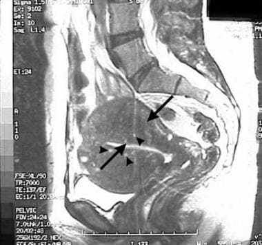
If the length of the entire uterus (including the cervix) is required (eg at preoperative evaluation), the sum of the total length of the uterine corpus (d1) and the cervical length should be reported d1 is calculated as the sum of the fundal length (from the fundal serosal surface of the uterus to the fundal tip of the endometrial cavity) and the endometrial cavity length (from theAdenomyosis was diagnosed when a poorly defined area of abnormal echotexture (decreased or increased echogenicity, heterogeneous echotexture, myometrial cysts) was present in the myometrium All endovaginal US findings were correlated with those from histologic examination Fig 1291 (A) Transvaginal ultrasound image shows an enlarged uterus with heterogeneous echotexture of the myometrium(B) Sagittal T2weighted image of the pelvis shows focal thickening of the junctional zone (arrow)There are T2 hyperintense foci seen in the thickened junctional zone Coronal T2weighted image of the pelvis shows a similar finding as


Www Aium Org Misc Soundjudgment6 Pdf


Cyberleninka Org Article N Pdf
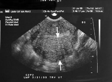
Dr Daniel J Bell and Dr Yuranga Weerakkody et al Coarsened hepatic echotexture is a sonographic descriptor used when the uniform smooth hepatic echotexture of the liver is lost This can occur due to a number of reasons which include conditions that cause hepatic fibrosis 1 cirrhosis hemochromatosisAs in the study by Reinhold et al , we found the most common sonographic finding in patients with diffuse adenomyosis to be a uterus with a diffuse heterogeneous echotexture Myometrial cystic spaces were seen in only three cases in our study These spaces correlated with fluidfilled dilated endometrial glands in the myometrium on histologic examination of the surgical specimenFrom this, practitioners can ensure that suspicious sonographic findings of uterine sarcomas, including a largesize, heterogeneous echotexture, central cystic change or necrosis, and hypervascularity, are recognized to facilitate further evaluation


Radiology Charts


The Normal Uterus
The uterine arteries reach the uterus at the level of the cardinal ligaments and divide into ascending and descending branches that travel within the layers of the broad ligament along the lateral wall Sonographically the normal myometrium has a medium echogenicity, less than the endometrium, with a granular echotexture The myometrium can be divided into three layersCirrhosis of the liver is a disease due to The echotexture of the right testicle is heterogeneous when compared to the normal homogenous echotexture of the left testicle The hypoechoic (dark) regions within the testicle represent areas of necrosis Note also the large complex hydrocele surrounding the necrotic right testicle with multiple septations, which is reactive in the setting of testicular torsion Doppler


Www Glowm Com Pdf Ultrasound In Obstetrics And Gynecology Chapter11 Pdf


Adenomyosis A Sonographic Diagnosis Radiographics
Ultrasound of the uterus with adenomyosis may reveal the organ to be of normal size or enlarged The echo texture of the affected regions is different from that of normal myometrium ; Grayscale ultrasound image of the uterus (a) demonstrates a heterogeneous uterine echotexture with poorly defined masses (arrow, (a)) The endometrial stripe is thickened (clippers, (a)) and measures up to 30 cm Color Doppler image (b) shows a soft tissue mass within the posterior wall of the urinary bladder (arrow, (b)) with vascularityIn some cases, the sole clue to the presence of leiomyoma on US is an irregularly enlarged uterus with distortion or obscuration of the endometrial stripe 6 When seen as discrete lesions, leiomyomas may appear hyperechoic or hypoechoic, homogeneous, or heterogeneous Regardless of their echogenicity or echotexture, shadowing and attenuation are the hallmarks of leiomyoma


Diseases Of The Uterus Radiology Key


What Are The Most Reliable Signs For The Radiologic Diagnosis Of Uterine Adenomyosis An Ultrasound And Mri Prospective Sciencedirect
Heterogeneous uterus Heterogenous uterus is a medically sonographic terminology It says how your uterus look under sonographic waves That is not a pathology in itself It says how your uterus look under sonographic waves


Www Glowm Com Pdf Ultrasound In Obstetrics And Gynecology Chapter11 Pdf


Adenomyosis And Ultrasound The Role Of Ultrasound And Its Impact On Understanding The Disease Obgyn Key


What Are The Most Reliable Signs For The Radiologic Diagnosis Of Uterine Adenomyosis An Ultrasound And Mri Prospective Sciencedirect


A Woman With Urinary Incontinence Nejm Resident 360 Meta Property Twitter Image Content Resident360files Nejm Org Image Upload C Fit F Auto H 1 W 1 V U8buf4o8mgdxgmfcczjk Png Meta Property Og Image Content


Retroverted Uterus Wikipedia


Adenomyosis A Sonographic Diagnosis Radiographics


Uterine Carcinosarcoma A Case Report And Literature Review


Imaging Of The Female Pelvis Through The Life Cycle Radiographics


Adenomyosis Common And Uncommon Manifestations On Sonography And Magnetic Resonance Imaging Chopra 06 Journal Of Ultrasound In Medicine Wiley Online Library


Sonohysterography Principles Technique And Role In Diagnosis Of Endometrial Pathology


Pelvic Imaging In Reproductive Endocrinology Oncohema Key


What Are The Most Reliable Signs For The Radiologic Diagnosis Of Uterine Adenomyosis An Ultrasound And Mri Prospective Sciencedirect


Adenomyosis And Its Impact On Fertility


Adenomyosis A Sonographic Diagnosis Radiographics


Pelvic Usg Uterus And Ovaries Radiology Key


Radiology Charts


When Would You Suspect Fibroid Uterus


Scielo Brasil Radiological Findings Of Uterine Arteriovenous Malformation A Case Report Of An Unusual And Life Threatening Cause Of Abnormal Vaginal Bleeding Radiological Findings Of Uterine Arteriovenous Malformation A Case Report


Adenomyosis Radiology Reference Article Radiopaedia Org


Www Aium Org Misc Soundjudgment6 Pdf


Gray Scale Transvaginal Us Demonstrating Uterus Increased In Volume Download Scientific Diagram


1


Pelvic Usg Uterus And Ovaries Radiology Key


Diffuse Uterine Adenomyosis Radiology Reference Article Radiopaedia Org


Adenomyosis Body Imaging Teaching Files Uw Radiology


Www Glowm Com Pdf Ultrasound In Obstetrics And Gynecology Chapter11 Pdf


Radiology In The Diagnosis Staging And Management Of Gynecologic Malignancies Glowm


Uterus Adenomyosis Ultrasound


The Uterus Myometrium Endometrium


Figure 3 Gestational Trophoblastic Disease A Multimodality Imaging Approach With Impact On Diagnosis And Management


Diseases Of The Uterus Radiology Key


Ultrasonography Of Uterine Leiomyomas Sciencedirect


The Assessment Diagnosis And Causes Of Endometrial Cancer Empowered Women S Health


Q Tbn And9gcq4n9kjisge1c2im3e Dttnlw8eipza2kkfxe6rmcfb2il5dfe6 Usqp Cau


Quantitative Analysis Of Ultrasound Images For Computer Aided Diagnosis


Adenomyosis Imaging Practice Essentials Magnetic Resonance Imaging Ultrasonography


Radiology Charts


Www Dsjuog Com Doi Dsjuog Pdf 10 5005 Jp Journals 1594


Diseases Of The Uterus Radiology Key


Adenomyosis Imaging Practice Essentials Magnetic Resonance Imaging Ultrasonography


Academic Oup Com Humupd Article Pdf 4 4 337 Pdf


Http Eknygos Lsmuni Lt Springer 295 61 100 Pdf


The Normal Uterus


Adenomyosis A Sonographic Diagnosis Radiographics


Venetian Blind Shadowing On Ultrasound Semantic Scholar


Uterine Adenomyosis Endovaginal Us And Mr Imaging Features With Histopathologic Correlation Radiographics


Adenomyosis Common And Uncommon Manifestations On Sonography And Magnetic Resonance Imaging Chopra 06 Journal Of Ultrasound In Medicine Wiley Online Library


Uterine Arteriovenous Malformation Radiology Case Radiopaedia Org
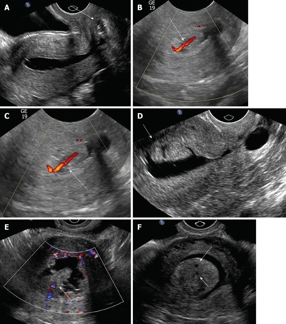


Sonohysterography Principles Technique And Role In Diagnosis Of Endometrial Pathology
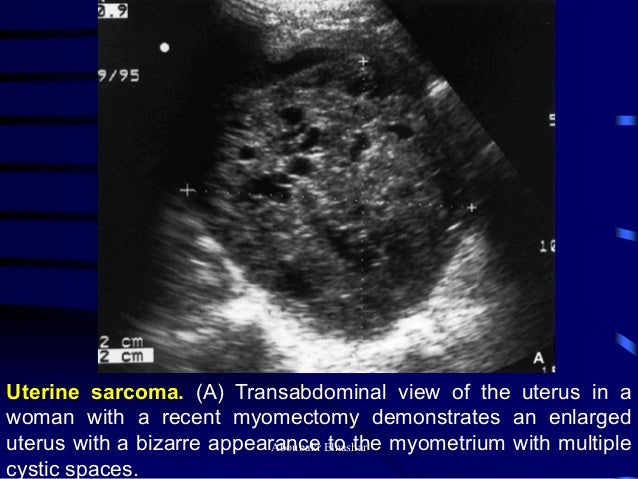


Ultrasonography Of The Uterus


Sonography Of Uterine Myometrial Disorders Youtube


Adenomyosis Radiology Case Radiopaedia Org


Adenomyosis Hyperechoic Nodules Top Left Sagittal Section Through Download Scientific Diagram


Adenomyosis Predominantly Hypoechoic Sagittal Section Through A Download Scientific Diagram


Adenomyosis Common And Uncommon Manifestations On Sonography And Magnetic Resonance Imaging Chopra 06 Journal Of Ultrasound In Medicine Wiley Online Library
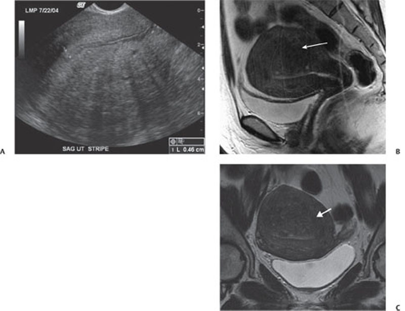


129 Adenomyosis Radiology Key


Ultrasonography Of Uterine Leiomyomas Sciencedirect
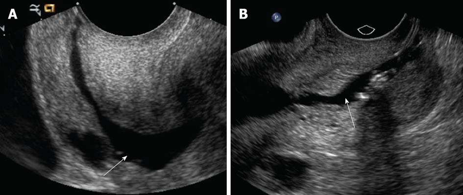


Sonohysterography Principles Technique And Role In Diagnosis Of Endometrial Pathology


Adenomyosis A Sonographic Diagnosis Radiographics


Imaging The Cervix Part I Mycme


Abnormally Thickened Endometrium Differential Radiology Reference Article Radiopaedia Org


Imaging Of The Female Pelvis Through The Life Cycle Radiographics
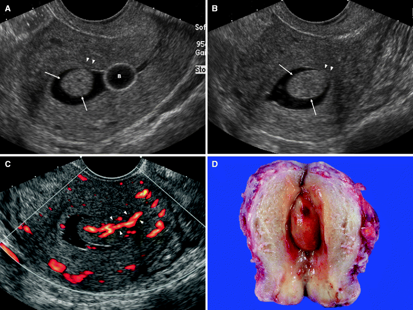


Illustrations Springerlink


Ultrasonography Of Uterine Leiomyomas Sciencedirect
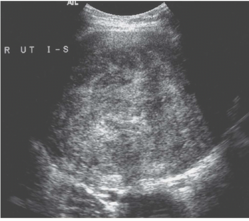


Diseases Of The Uterus Radiology Key


Radiology Charts


Adenomyosis A Sonographic Diagnosis Radiographics


Adenomyosis Common And Uncommon Manifestations On Sonography And Magnetic Resonance Imaging Chopra 06 Journal Of Ultrasound In Medicine Wiley Online Library


Oblique View Of The Uterus With The Probe Tilted Towards The Right Download Scientific Diagram


The Accuracy Of Transvaginal Ultrasound And Uterine Artery Doppler In The Prediction Of Adenomyosis Sciencedirect


What Does A Homogeneous Myometrium Mean Quora


Radiology Charts
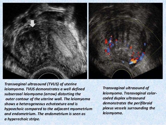


Presentation1 Pptx Radiological Imaging Of Uterine Lesions


Adenomyosis Common And Uncommon Manifestations On Sonography And Magnetic Resonance Imaging Chopra 06 Journal Of Ultrasound In Medicine Wiley Online Library
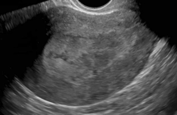


Recent Updates In Female Pelvic Ultrasound Springerlink


Adenomyosis Radiology Reference Article Radiopaedia Org
コメント
コメントを投稿